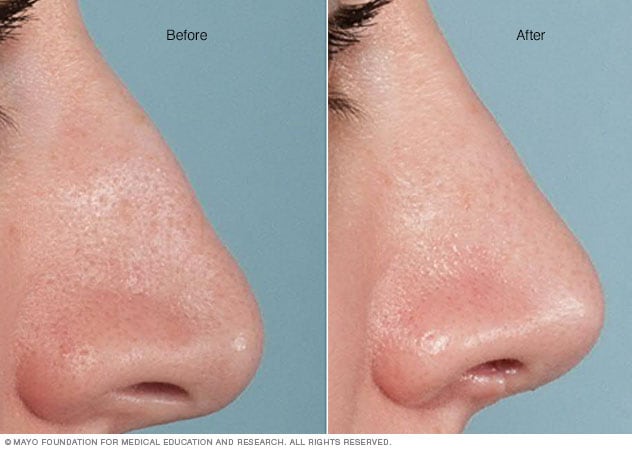
Get This Report on Rhinoplasty Surgeon Austin
Table of Contents5 Easy Facts About Rhinoplasty Surgeon Austin ExplainedFacts About Austin Rhinopasty Surgeon Uncovered
The design template is turned 180 degrees and put over the distal (far) portion of the axis of the skin flap; the surgeon describes it with a surgical marker. The overview markings are continued proximally and parallel to the main axis, preserving a 2-cm width for the proximal flap. Without using an injection of anaesthetic epinephrine, the flap is incised (cut), and the distal one-half rises in between the frontalis muscle and the subcutaneous fat.The dissection continues toward the eyebrow and the glabella (the smooth prominence between the eyebrows) until the skin flap is adequately mobile to enable its unwinded transposition upon the nose. Under loupe magnification, the distal portion of the forehead flap is de-fatted, to the subdermal plexus. Yet, the fat-removal must be conservative, especially if the client is either a tobacco smoker or a diabetic, or both, since such health aspects negatively impact blood circulation and tissue perfusion, and thus the prompt and right healing of the surgical scars to the nose.

Surgical technique the septal mucosal flap The cosmetic surgeon cuts the anteriorly based septal mucosal tissue-flap as commonly as possible, and after that launches it with a low, posterior back-cut; however just as needed to allow the rotation of the tissue-flap into the nasal injury. The surgeon measures the dimensions (length, width, depth) of the nasal wound, and then marks them upon the nasal septum, and, if possible, incorporates an additional margin of 35 mm of width to the injury measurements; furthermore, the base of the mucosal tissue flap should be at least 1.
The cosmetic surgeon then makes two (2) parallel incisions along the flooring and the roof of the nasal septum; the incisions converge anteriorly, towards the front of the nasal spine. Using an elevator, the flap is dissected in a sub-mucoperichondrial aircraft. The (far) distal look here edge of the flap is cut with a right-angle Beaver blade, and after that is transposed into the injury.
A technical variant of the septal mucosal flap method is the Trap-door flap, which is utilized to rebuild one side of the upper half of the nasal lining. It is emplaced in the contralateral nasal cavity, as a superiorly based septal mucosal flap of rectangular shape, like that of a "trap-door".
The surgeon raises the flap of septal mucosa to the roof of the nasal septum, and after that traverses it into the contralateral (opposite) nasal cavity through a slit made by getting rid of a little, narrow part of the dorsal roofing of the septum. Afterwards, the septomucosal flap is extended across the wound in the mucosal lining of the lateral nose - austin rhinopasty surgeon.
The 45-Second Trick For Austin Rhinopasty Surgeon
I. Partial-thickness defects A partial-thickness defect is a wound with adequate soft-tissue protection of the underlying nasal skeleton, yet is too large for primary intent closure, with sutures. Based upon the area of the injury, the cosmetic surgeon has 2 (2) choices for fixing such an injury: (i) healing the injury by secondary objective (re-epithelialisation); and (ii) healing the injury with a full-thickness skin graft (rhinoplasty austin tx).
In the event, bigger nasal wounds (defects) do successfully heal by secondary objective, but do present 2 downsides. First, the resultant scar frequently is a large patch of tissue that is aesthetically inferior to the scars produced with other nasal-defect correction techniques; however, the skin of the medial canthus is an exception to such scarring.
Yet, nasal correction with a skin graft harvested from the client's neck is not suggested, because that skin is low-density pilosebaceous tissue with really few hair follicles and sebaceous glands, therefore differs from the oily skin of the nose. The technical benefits of nasal-defect correction with a skin graft are a short surgery time, a basic rhinoplastic method, and a low incidence of tissue morbidity.
